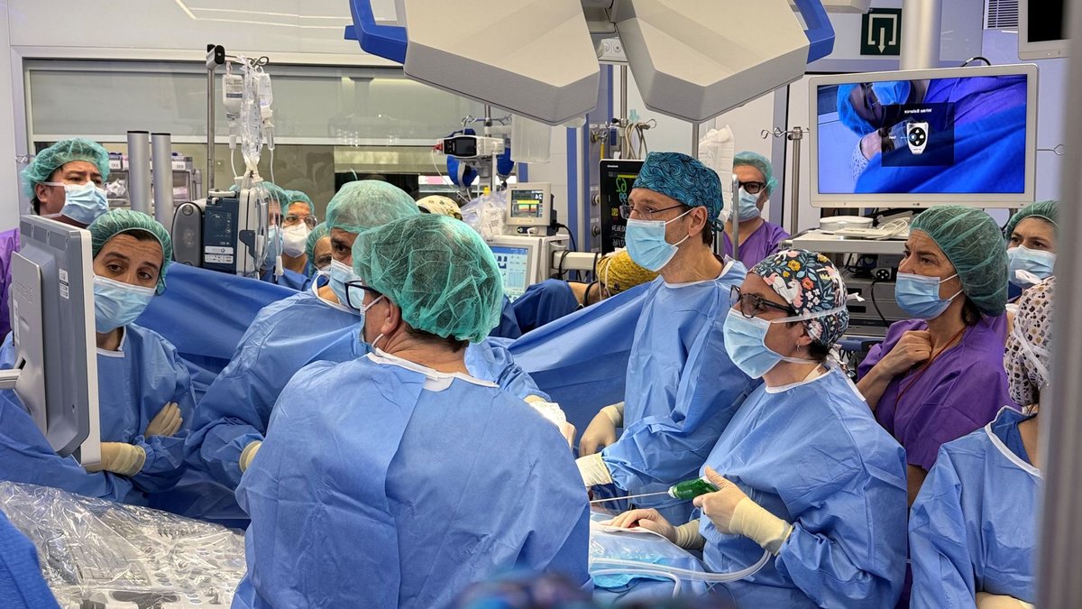
A team of specialists has performed Europe’s first operation on a foetus whose entire intestine had developed outside its body, according to reports from SJD Barcelona Children’s Hospital. The medical team decided to perform foetal surgery at 28 weeks’ gestation to prevent irreversible damage to the baby. The procedure, which took place on 10 February, lasted two and a half hours and was carried out by a team comprising specialists in foetal medicine and surgery, paediatric surgery, anaesthesiology and neonatology.
The foetus had gastroschisis, a congenital malformation in which the baby’s intestines develop outside the body through an opening in the abdominal wall, located to the right of the umbilical cord. The opening does not close properly during pregnancy, leaving the intestines exposed to amniotic fluid. This exposure can cause inflammation, loss of blood supply, and necrosis, with serious consequences for the baby after birth. The healthcare professionals monitoring the pregnancy of the mother, a Colombian 20-year-old woman, detected the foetal malformation during a routine ultrasound scan in the 20th week of gestation and referred her to BCNatal, a leading centre in obstetrics and foetal medicine.
Related: A Deeper Look at Multiple Myeloma in the Middle East
After evaluating the various options and consulting the case with the ethics committee, the medical team proposed a foetal surgery that has so far been performed only on very rare occasions in Colombia and the United States, but never in Europe. The surgeons made an incision in the mother’s abdomen to access the uterus; they then repositioned the foetus to allow access to its abdomen and performed the procedure laparoscopically. To do this, they inserted four trocars into the amniotic sac and, after draining the fluid and introducing gas to create space within the abdomen that would allow them to see and operate safely, they carefully repositioned the intestine within the abdominal wall.
Upon completion, they closed the incision in the abdomen. This is a procedure that requires a high degree of precision, given that the foetus weighed 700 grams, the intestine was 80 centimetres long and had a diameter of 3 to 15 mm, and had to be reinserted through a puncture measuring just 1.5 centimetres. In the days following the procedure, ultrasound scans confirmed that the inflammation in the intestine was subsiding and that it was returning to its normal appearance.
This case is a rare success in a field where outcomes are often unpredictable. While the medical team handled a complex anatomy with limited space, the precise reinsertion of the bowel suggests that the procedure, while technically demanding, can stabilize a pregnancy that would otherwise be at high risk of miscarriage or severe neonatal complications.
Related: Turning awareness into cancer appointments
Normal development after birth
The pregnancy continued and the baby was delivered by caesarean section at 34 weeks’ gestation. Although the baby was underweight for its gestational age, the outcome was very favourable and, after a short stay in hospital, the baby was discharged. The baby is currently feeding normally.
Gastroschisis occurs in between 2 and 3 out of every 10,000 births, which translates to 120 cases annually in Spain and 2,400 in Europe. Foetal surgery would only be indicated in 10% of these cases, making this intervention a highly specialized procedure reserved for specific medical circumstances. The successful operation marks a significant step in the management of this condition in Europe.



Leave a Reply