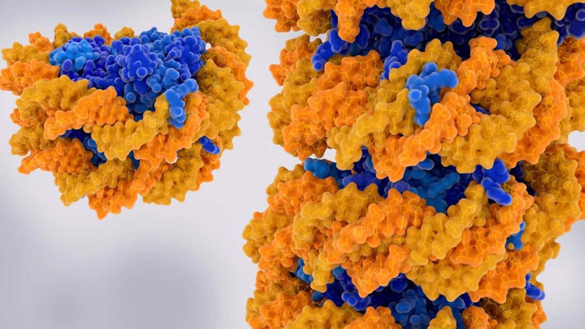
A deep-learning AI tool can now identify aging patterns in blood stem cells by studying how DNA is organized within the cell nucleus. Scientists at the Bellvitge Biomedical Research Institute and the Barcelona Supercomputing Center created ChromAgeNet, a model trained on three-dimensional microscopy images of hematopoietic stem cells (HSCs). Their findings, published in Aging Cell, demonstrate that chromatin architecture, the spatial arrangement of DNA and proteins inside the nucleus, holds enough information for AI to differentiate between young and aged cells, even when visual distinctions are minimal.
HSCs play an essential role in sustaining blood and immune cell production, but their efficiency diminishes over time. While most aging research has centered on molecular indicators like DNA methylation, ChromAgeNet instead focuses on the physical structure of chromatin. The team trained the model using DAPI-stained HSC nuclei from young and aged mice, allowing it to learn from full 3D image stacks rather than relying on pre-selected features. This method enhanced precision by evaluating entire nuclear structures instead of isolated areas.
The researchers applied explainable AI methods to pinpoint which features influenced the model’s predictions. Critical indicators included chromatin entropy, peripheral heterochromatin accumulation, and chromatin condensates. In younger cells, informative regions tended to cluster near the nuclear envelope, whereas aged cells exhibited more scattered patterns, indicating greater chromatin disorganization.
Read Also: New blood test accurately distinguishes bacterial and viral infections
Next, they applied ChromAgeNet to aged HSCs exposed to compounds that interfere with Rho GTPase signaling or chromatin regulation. Some treatments adjusted the cells’ AI-generated “youthful scores” toward those of younger HSCs, with chromatin-modifying agents producing the most significant shifts. This suggests the tool could function as a screening platform for rejuvenation studies, as DAPI staining remains a low-cost and widely adopted microscopy technique.
Yet, a visually youthful nucleus does not confirm functional rejuvenation. The authors stress that biological validation is required before ChromAgeNet scores can verify true cellular rejuvenation. Currently, the model operates as a preclinical concept, developed in mouse cells where chronological age serves as a proxy for biological aging. Differences in imaging protocols and cell variability will need further validation across laboratories and microscopy systems.
Adapting the model to human HSCs introduces additional complexity, as mouse and human chromatin structures differ. The team anticipates modifications will be necessary before applying ChromAgeNet to human samples. The broader opportunity lies in establishing a measurable phenotype for drug development, one capable of assessing not only a cell’s apparent age but whether interventions meaningfully alter its aging trajectory.




Leave a Reply